Investigation of metal 3D printed materials
Metal 3D printed samples were prepared by Metalloexpert ltd. from Markedforged 17-4PH stainless steel materials. 17-4PH stainless steel commonly because it offers high strength and hardness along with excellent corrosion resistance.
The morphology of Metalloexpert’s sample was investigated by our Zeiss Sigma 300 field emission electron microscope. Thanks to Zeiss Gemini technology we able to use extra low acceleration voltage to minimize charge up effects and increase surface sensitivity. It also allows SEM investigation of nonconductive materials without extra coating. SEM images were captured from each processing phases: filament, green-, brown- and sintered parts. In the filament metal beads with size 0,5-7 µm are evenly embedded into polymer matrix.
Filament


Green Part
After the printing (green part) the matrix appears denser and smoother. This change is most probably caused by the shearing of the extrusion.


Brown Part
Brown parts are prepared by partial removal of the polymer matrix from green parts. SEM images confirm the success of the washing process: there are much less polymer can be detected between the metal particles in case of brown part.


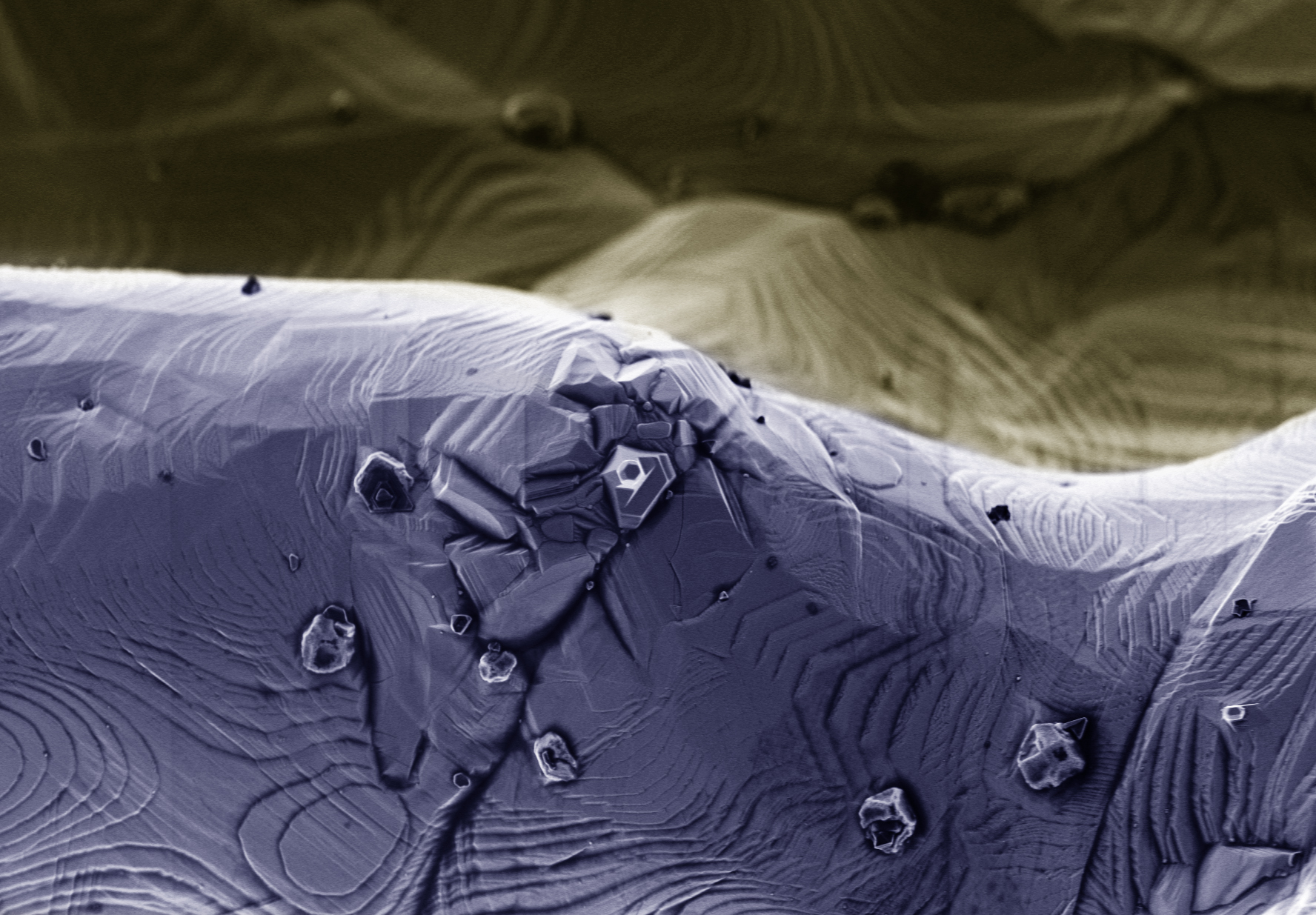
Sintered Part
The morphology of the sintered part is extremely different from previous steps. The particles are transformed into bulk metal. Its pristine surface displays well-defined steps and terraces with copper-rich crystals.


Results
Images with lower magnification from the outer area give us information about the printing quality. However, some holes can be seen around printing errors, the most part of the sintered material is even and non-porous.

Printing defects
